Please contribute your submission via https://mc03.manuscriptcentral.com/lam. Please mark that it is a contribution to Special Issue on Light and Cancer: Tools for Diagnostics and Theranostics in the cover letter upon submission.
Submission deadline: 31st December, 2024
Introduction to the Special Issue:
The goal of this special issue is to present the most exciting developments in the field of light technologies for the earlier detection, classification and treatment of cancer. The state-of-the-art of the problem, as well as the broad and insightful prospects of this urgent field of technology, gathering the advanced achievements of international research groups and demonstrating scientific breakthroughs and practical prospects for the use of light in cancer research and treatment. Special attention will be paid to multimodal approaches that guarantee high accuracy of diagnosis and treatment, where optical technologies [1] can become a powerful addition to high-tech modern imaging modalities such as ultrasound, computed tomography, magnetic resonance imaging and positron emission tomography. Most optical technologies offer great potential for such a combination with the possibility of using fiber-optics in clinical settings, including endoscopy.
Currently, optical technologies themselves also provide multimodality in cancer research through the use of various combinations of techniques with different spatial resolution and depth of sensing, which serve as instruments of “optical biopsy”, namely functional near-infrared spectroscopy (fNIRS), diffusion optical tomography (DOT), hyperspectral imaging (HSI), fluorescence lifetime imaging (FLIM), photoacoustic (PAT) and acoustooptic (AOT) tomographies, multiphoton imaging, optical coherence tomography (OCT), confocal and light sheet microscopies, as well as Raman-based techniques (V.V. Tuchin, J. Popp, and V.P. Zakharov (Eds.), Multimodal optical diagnostics of cancer, Basel: Springer Nature Switzerland AG, 2020). In turn, approaches to enhancing Raman scattering, such as surface-enhanced Raman scattering (SERS), coherent anti-Stokes Raman scattering (CARS) and stimulated Raman scattering (SRS), can significantly improve the molecular fingerprinting of cancerous tissue for malignancy diagnostics and classification on molecular level. OCT also offers a diversity of powerful techniques for characterizing cancerous tissues, such as polarization-sensitive OCT (PS-OCT), which allows complementary images to be obtained for the initial polarization and cross-polarization states (CP-OCT), or images for all elements of the complete Mueller matrix or combinations thereof. There are many other OCT tools: label-free OCT angiography (OCTA), OCT-based elastography (OCE), line-field confocal OCT (LC-OCT), Doppler OCT (DOCT), speckle-resolved OCT and others. Current multiphoton imaging systems for in vivo studies use second (SHG) and third (THG) harmonic generation techniques, two-photon microscopy (2PM) and three-photon microscopy (3PM), CARS and FLIM modalities.
Potential topics include, but are not limited to:
Optical imaging and spectral analysis of precancer and early cancer tissues
Multimodal spectral diagnostics and optical imaging
Optical properties of various types of cancerous tissues
fNIRS for cancer diagnostics and monitoring of therapy
Light-induced autofluorescence analysis
UV fluorescence endoscopy applications for cancer diagnosis
Diffuse reflectance spectroscopy for the diagnosis of skin and oral tumors
FLIM in probing metabolism in cancer
Raman spectroscopy, SERS, CARS, SRS
Multiphoton imaging, 2-PM, 3-PM, SHG, THG
OCT, multimodal OCT
Hyperspectral imaging devices
Fine-needle optical probes
Multimodal fiber-optical probes
Terahertz (THz) spectroscopy and imaging for label-free diagnosis of malignancies
Tissue optical clearing
Optical studies of blood, extracellular vesicles for diagnosis, monitoring, and prediction of cancer
Spectral analysis of breath air samples as a diagnostic tool
Multimodal cancer imaging
Malignancies with different nosology and localization
Devices for screening, early detection at the point-of-care, treatment monitoring, and image-guided intervention
Dermatoscopy imaging
Biopsy guidance and real-time thick tissue histology
Sensitivity and specificity of cancer diagnosis
Data analysis and multivariate statistics, software algorithms based on machine learning
This issue is co-edited by:

Prof., Dr. Sc. Valery V. Tuchin, Saratov State University, Russia
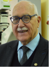
Valery V. Tuchin is the corresponding member of the Russian Academy of Sciences, Head of the Department of Optics and Biophotonics and Director of the Science Medical Center of Saratov State University. He is also works with several other research centers of RAS and universities. His research interests include biophotonics, tissue optics, drug delivery, and phototherapy. He is the Honored Scientist of Russia, Fellow of SPIE and OPTICA, Distinguished Professor of Finland, recipient of SPIE Educator Award, Joseph W. Goodman Book Writing Award (OPTICA/SPIE), Michael S. Feld Biophotonics Award (OPTICA), Chime Bell Award of the Chinese province of Hubei, the D.S. Rozhdestvensky Medal and the S.I. Vavilov Medal of the Russian Optical Society, and the A.M. Prokhorov Medal of the Russian Academy of Engineering Sciences.

Juergen Popp, Friedrich-Schiller University Jena and Leibniz Institute of Photonic Technology Jena, Germany
Since 2002 Juergen Popp is holds a chair for Physical Chemistry at the Friedrich-Schiller University Jena, Germany. Since 2006 he is also the scientific director of the Leibniz Institute of Photonic Technology, Jena. Juergen Popp is a world leading expert in Biophotonic / optical health technology research covering the complete range from photonic basic research towards translation into clinically applicable methods. He has published more than 990 journal papers, has been named as an inventor on 15 patents and has given more than 200 invited talks on national and international conferences (among them more than 50 keynote/plenary lectures). He received several awards like e.g. the 2016 Pittsburgh Spectroscopy Award, the Kaiser-Friedrich-Forschungspreis 2018, the third price of the Berthold Leibinger Innovationspreis in 2019 or in 2022 he became part of The Photonics100. Furthermore, he received two honorary doctorate degree: 2012 from the Babes-Bolyai University in Cluj-Napoca, Romania and very recently 2023 from the University at Albany – State University of New York (USA).

Valery P. Zakharov, Samara National Research University, Samara, Russia
Valery P. Zakharov is a Professor and Chair of Laser and Biotechnical Systems Department, Head of Photonics Laboratory at Samara National Research University. His research interests include laser medicine, biophotonics, multimodal biomedical imaging, and application of fluorescence and Raman-techniques for biomedical diagnostics. He has published more than 350 publications in peer-reviewed journals, has several patents and yearly has given invited talks on national and international conferences. He has been awarded Honorary Worker of Education of Russian Federation, Samara Award for Biophotonics innovations.


 Email
Email RSS
RSS